Digital radiography, for students and faculty.

For dental, dental hygiene and dental assisting programs: the sensor sizes at true scale, what the resolution figures mean, how to handle and care for a sensor, and real X-rays to compare imaging algorithms on.

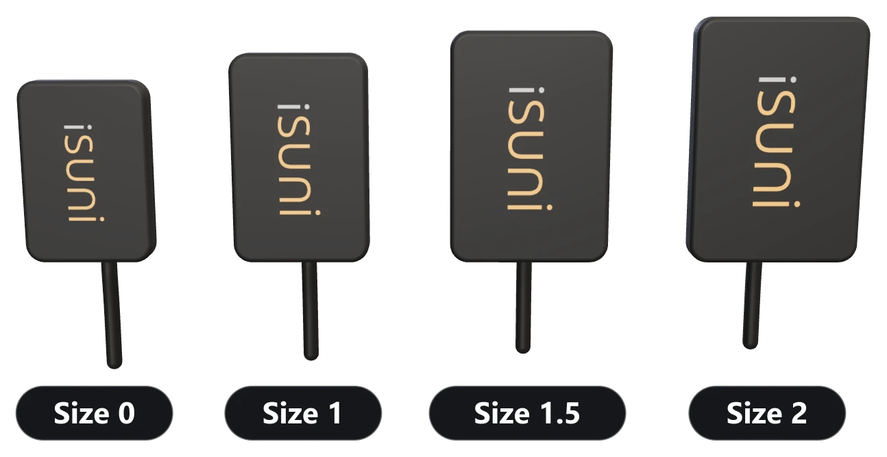
iSuni intraoral sensors in sizes 0, 1, 1.5 and 2, side by side to scale.

Used by schools and examiners

Schools, hospitals and examining boards that use iSuni sensors.

All third-party trademarks and logos shown are the property of their respective owners.

Sensor sizes, at true scale

Most clinics need one or two sizes, not four. Choose a size to see its plate, its active image area and its figures. How to choose a sensor size

Choose a sensor size
011.52

At true scale: each square is 1 cm

Size
0
Outside
23.4 × 33.6 mm, 4.5 mm thick
Active image area
18 × 24 mm
Image
900 × 1200 pixels (1.08 MP)
Pixel pitch
20 µm
Resolution
20 lp/mm measured, 25 lp/mm theoretical
Size
1
Outside
25.0 × 38.5 mm, 4.5 mm thick
Active image area
20 × 30 mm
Image
1000 × 1500 pixels (1.50 MP)
Pixel pitch
20 µm
Resolution
20 lp/mm measured, 25 lp/mm theoretical
Size
1.5
Outside
29.2 × 42.5 mm, 4.5 mm thick
Active image area
24 × 33 mm
Image
1200 × 1650 pixels (1.98 MP)
Pixel pitch
20 µm
Resolution
20 lp/mm measured, 25 lp/mm theoretical
Size
2
Outside
31.0 × 45.0 mm, 4.5 mm thick
Active image area
26 × 36 mm
Image
1300 × 1800 pixels (2.34 MP)
Pixel pitch
20 µm
Resolution
20 lp/mm measured, 25 lp/mm theoretical

Measured or theoretical?

Spec sheets often quote only the theoretical figure. Choose a pattern to see what 20 µm pixels can record. Sensor resolution, explained

Choose a pattern

10 lp/mm. Each line covers more than two pixels: recorded clearly.

20 lp/mm, measured. Still separate lines on a perfect grid. In a real sensor, light spreading in the scintillator softens them: 20 lp/mm is the finest pattern the iSuni sensor resolves.

25 lp/mm, the pixel limit. One pixel per line, and only when the pattern lines up exactly with the pixels: the theoretical maximum of a 20 µm pixel.

35 lp/mm, beyond it. Finer than the pixels can follow: they record a false, coarser pattern instead. No 20 µm sensor can show this.

  • The pattern
  • What the pixels record (20 × 20 µm)
  • Illustration on a perfect 20 µm grid, not a measurement.

Placing and looking after a sensor

The full sensor care routine

  • Rounded, chamfered corners help with positioning, on a 4.5 mm plate.
  • Positioners and sleeves made for digital sensors of that size; most clinics keep one holder kit per size.
  • A new barrier sheath for every patient.
  • Mind the cable: never pull the sensor out by its cable, bend it sharply, roll a chair over it or let a patient bite it.
  • Wipe, never soak: the head and cable with 70% isopropyl alcohol, the USB plug kept dry, and never autoclaved or heat-sterilized.
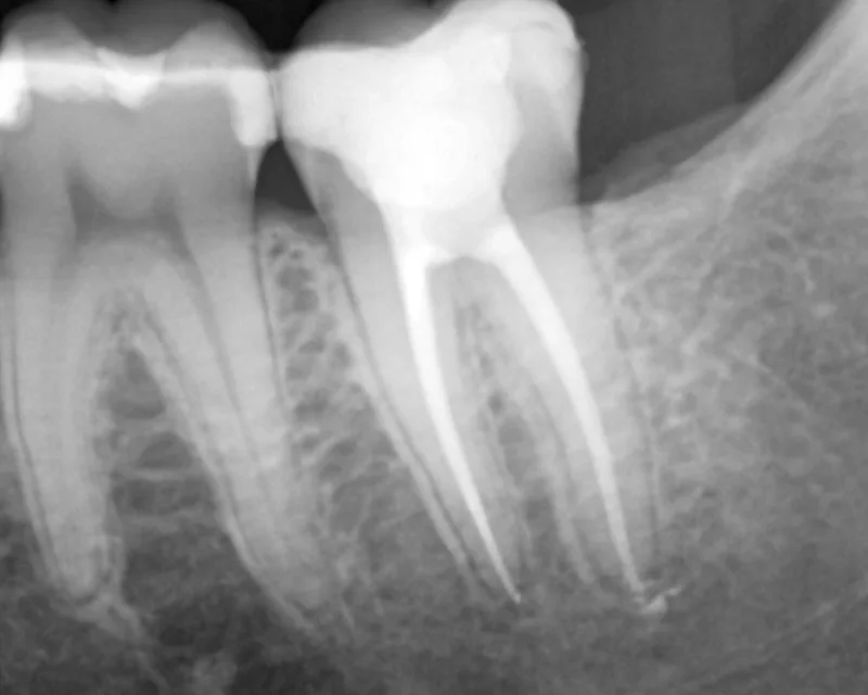
A periapical X-ray from an iSuni sensor with the SuniVision™ Clear imaging algorithm.

Real X-rays, two algorithms

SuniVision™ Studio shows real periapical X-rays from iSuni sensors, as taken and with each of the two SuniVision™ imaging algorithms, Clear and Detail. Drag across an image to compare.

Open SuniVision™ Studio

Equipping a teaching clinic?

Tell us about your program, your operatories and your imaging software, and we’ll reply with a quote.

Your quote

Nothing in your quote yet. Add a sensor size, a scanner or the C8 camera, then request one quote for everything.

Subtotal

List prices in Canadian dollars, before tax. iSuni confirms your price in the quote.

Request this quote

Chat with us (opens in a new tab)